

Key facts about melanoma
Melanoma is a cancer that develops from melanocytes, the cells responsible for producing skin pigment. It is less common than other types of skin cancer, but it can grow deeper into the skin and spread to the lymph nodes and other organs if it is not diagnosed early.
The most common sign is the appearance of a new mole or spot, or a change in an existing lesion. Asymmetry, irregular borders, multiple colors, and progressive growth increase suspicion. However, melanoma can also be small, have a uniform color, or appear as a pink, reddish, or skin-colored lesion.
Melanoma can develop anywhere on the body, including the scalp, palms of the hands, soles of the feet, and under the nails. People with darker skin can also develop melanoma, and lesions on the extremities and under the nails are proportionally more common in these patients.
The diagnosis is confirmed by biopsy. Dermoscopy helps dermatologists select suspicious lesions, but photographs, apps, and blood tests cannot replace microscopic examination of the tissue.
When detected while still superficial and confined to the skin, melanoma can often be cured with surgery. In cases with a higher risk of recurrence or spread, immunotherapy, drugs targeting specific mutations, radiation therapy, or other cancer treatments may be necessary.
Any mole that is growing, changing shape or color, bleeding, developing a sore, or becoming very different from the others should be examined by a dermatologist.
Melanoma and Other Types of Skin Cancer
There are basically three major types of skin cancer:
- Basal cell carcinoma.
- Squamous cell carcinoma.
- Malignant melanoma.
Skin cancer is the most common type of cancer. Non-melanoma skin cancers, which mainly include basal cell carcinoma and squamous cell carcinoma, account for most cases. Melanoma is much less common but has a greater ability to invade deeper tissues and metastasize (spread to other parts of the body).
Ultraviolet radiation is the main cause of skin cancer. In addition to sunlight, this radiation is also emitted by tanning beds.
The effects of ultraviolet radiation on the skin are cumulative. This means that even after excessive sun exposure has stopped, skin changes may still appear years later.
What Is Melanoma?
Melanin is a pigment produced by melanocytes and is responsible for the color of our skin, eyes, and hair. When we are exposed to sunlight, melanocytes are stimulated to produce more pigment, causing the skin to darken.
Melanoma is a malignant tumor that develops from melanocytes, the skin cells responsible for producing melanin. Melanocytes are found mainly in the skin, but they are also present in the eyes, gastrointestinal tract, meninges, and the mucous membranes of the mouth and genital tract. This means that although the vast majority of melanomas are skin cancers, they can occasionally develop in the eyes or intestines, for example.
Although melanoma is much less common than basal cell and squamous cell carcinomas, it is the skin cancer with the greatest potential to spread. When diagnosed while still superficial and confined to the skin, surgery offers a high chance of cure. The risk increases as the tumor grows deeper or reaches the lymph nodes and other organs.
This article focuses mainly on cutaneous melanoma. Melanomas that arise in the eyes or mucous membranes are rare forms of the disease and have somewhat different biological characteristics, staging systems, and treatments.
Melanoma Causes and Risk Factors
People with fair skin, red or blond hair, light-colored eyes, freckles, and a tendency to sunburn have a higher risk of melanoma because they have less natural protection against ultraviolet radiation. This does not mean that people with darker skin are protected: melanoma can occur in people of any skin tone and, in these patients, lesions on the palms, soles, or under the nails deserve particular attention.
Having many moles, atypical moles, or a personal history of melanoma also increases the risk. The more moles a person has, especially if some differ in size, shape, or color, the more important regular skin examinations by a dermatologist become.
Sun exposure is the main risk factor for all types of skin cancer, including melanoma.
The pattern and cumulative amount of sun exposure throughout life are associated with the type of skin cancer a person may develop.
Non-melanoma skin cancers tend to occur in people with high cumulative sun exposure throughout life and appear mainly on the most sun-exposed areas of the skin, such as the face, hands, and forearms.
Melanoma, on the other hand, tends to occur in people who have less frequent but more intense sun exposure. One example is a fair-skinned person who receives excessive sun exposure while on vacation and develops painful, extensive sunburn.
These intermittent but intense exposures are particularly harmful when they occur during childhood and adolescence. People who have had more than five episodes of excessive sun exposure with significant sunburn have up to twice the risk of developing melanoma in adulthood.
Other risk factors for melanoma
- Indoor tanning: tanning beds emit carcinogenic ultraviolet radiation and increase the risk of melanoma. The risk rises with younger age at first use and with more frequent use. According to the World Health Organization, first using a tanning bed before the age of 35 is associated with an approximately 59% increase in melanoma risk.
- Family history: if a first-degree relative (a parent, sibling, or child) has had melanoma, you also have a greater genetic predisposition to developing the disease.
- Immunosuppression: people with weakened immune systems, such as patients with AIDS, organ transplant recipients, or people receiving chemotherapy, have a higher risk of developing melanoma.
- Living in areas with high ultraviolet radiation: the risk of sun-related melanoma is particularly high in people with fair skin. In people with darker skin, this risk is lower but not absent.
Note: acral melanoma, which occurs mainly on the palms, soles, and nails, is proportionally more common in people with darker skin and does not have a clear association with ultraviolet radiation.
How to Prevent Melanoma
Because at least two-thirds of melanoma cases are caused by excessive sun exposure, the main preventive measure against skin cancer is to reduce exposure to ultraviolet radiation.
Recommendations for sun and ultraviolet exposure:
- Avoid sun exposure severe enough to cause sunburn.
- Do not use tanning beds.
- Use sunscreen with an SPF of at least 30, ideally 50. Reapply it every two hours.
- Sunscreen does not replace the need to avoid prolonged sun exposure.
- Do not skimp on sunscreen. Many people use too little in an attempt to make the bottle last longer and therefore do not apply enough to provide adequate skin protection.
- Wear sunglasses with UV protection.
- Avoid sun exposure between 10 a.m. and 4 p.m.
- Limit deliberate sunbathing.
- Wear a hat when you expect to spend a long time in the sun.
- When working outdoors, wear lightweight clothing that covers your arms and legs.
Melanoma Symptoms
Melanoma can develop anywhere on the body, but it commonly appears in areas that have received intense sun exposure, such as the back, arms, legs, and face.
Melanoma can also develop in areas with little sun exposure, such as the soles of the feet, palms of the hands, and underneath the nails. Because melanocytes are also present in other parts of the body, melanoma can, less commonly, occur in the eyes, intestines, and genital tract.
The first sign is often a new dark spot on the skin or a change in the characteristics of an existing mole.
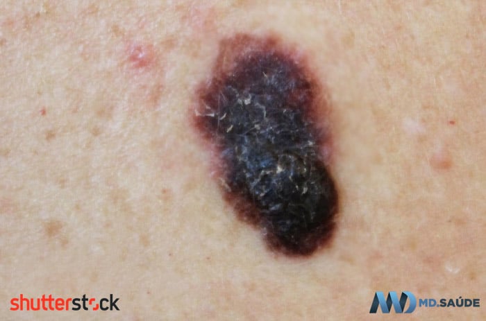
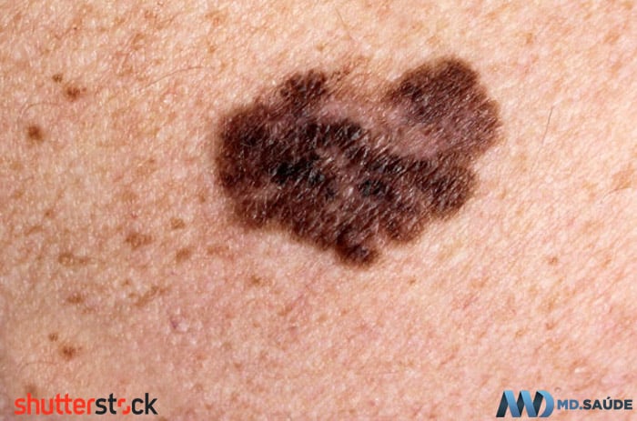
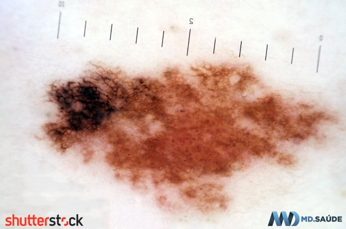
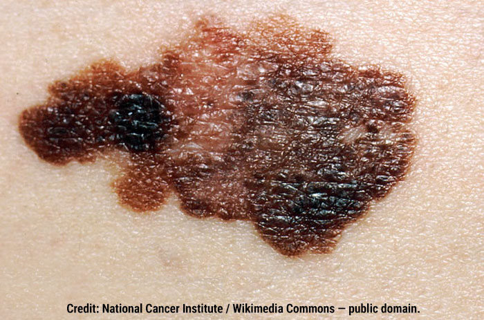
ABCDE
The ABCDE rule is a guide used to help doctors and patients identify moles and other skin lesions that should be evaluated by a dermatologist. Suspicious lesions may have the following characteristics:
A — Asymmetry: common moles usually have a symmetrical shape, typically oval or round. Melanomas are often asymmetrical.
B — Border: melanomas may have irregular, notched, blurred, or poorly defined borders. Benign moles usually have smoother, more clearly defined borders, although this criterion should not be assessed in isolation.
C — Color: melanomas often show variations in color within the same lesion, with areas that may be brown, black, red, blue, gray, or white.
D — Diameter: common moles are usually no larger than 6 mm (0.24 inches) in diameter. Therefore, lesions larger than 6 mm deserve attention. However, melanomas can also be smaller, especially in the early stages; a small mole should not automatically be considered benign simply because it measures less than 6 mm.
E — Evolving: moles that change in shape, color, size, or elevation over time should be considered suspicious. The development of bleeding, ulceration, persistent itching, or other symptoms also warrants medical evaluation.
Another useful approach is the so-called “ugly duckling” sign. In general, the moles on the same person tend to follow a similar pattern. A lesion that looks very different from the others in shape, color, size, or overall appearance deserves attention, even if it does not meet all the ABCDE criteria.
It is important to emphasize that most moles do not turn into melanoma, just as most melanomas do not develop from a pre-existing mole.
Some melanomas begin as flat lesions and later develop a raised area. Others, such as nodular melanoma, may appear from the beginning as a growing nodule. Whether a lesion is raised does not, by itself, indicate how deeply the tumor has grown, because many benign moles are also raised.
The ABCDE rule helps identify suspicious lesions, but it cannot confirm or rule out melanoma.
Melanoma Pictures
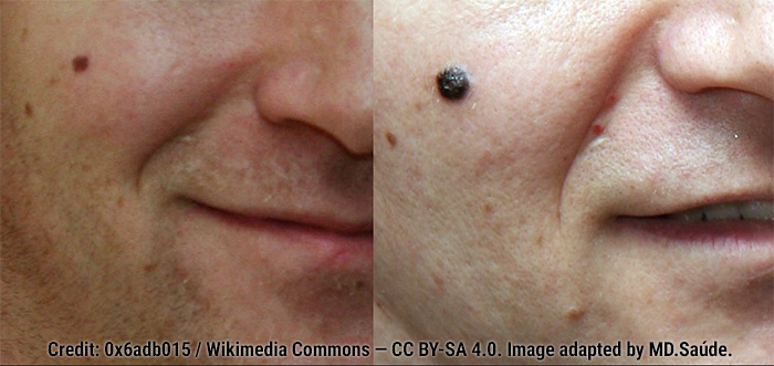
Melanomas That Do Not Follow the Classic Pattern
Not every melanoma is a dark mole. Amelanotic melanoma produces little or no melanin and may appear as a pink, reddish, or skin-colored lesion. Because it lacks the pigmentation typically associated with melanoma, it may be mistaken for a pimple, wart, sore, or another type of skin lesion.
A pink or reddish lesion that grows, changes in appearance, bleeds, or does not heal should be examined even if it is not brown or black.
Melanoma can also develop under a nail, a condition known as subungual melanoma. It may appear as a vertical dark streak, usually brown or black, that becomes wider or more irregular over time. In some cases, the pigment extends onto the skin surrounding the nail.
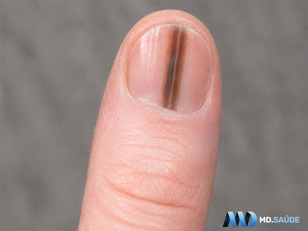
Not every dark streak in a nail is melanoma. Trauma, small areas of bleeding, medications, and natural pigmentation can also produce similar discoloration. However, a new, progressive, or irregular streak, particularly when it occurs in only one nail, should be evaluated by a dermatologist.
Melanoma Diagnosis
Diagnosis begins with a clinical examination of the suspicious lesion. The dermatologist evaluates features such as shape, borders, colors, size, elevation, and changes over time. The lesion is also compared with the patient’s other moles to identify any that look markedly different.
Dermoscopy is an examination performed with a device that magnifies the lesion and allows structures that cannot be seen with the naked eye to be visualized. It helps distinguish benign moles from suspicious lesions and helps determine which lesions should be removed, but dermoscopy alone cannot confirm a diagnosis of melanoma.
The definitive diagnosis is made by biopsy, in which the lesion is examined under a microscope. Whenever possible, the entire suspicious lesion is removed along with a small margin of surrounding skin. If the lesion is very large or located in an area where complete initial removal would be difficult, the doctor may remove only a representative portion for analysis.
In addition to confirming or ruling out melanoma, the biopsy report provides important information such as Breslow thickness, the presence of ulceration, and whether the surgical margins are involved. These findings help determine the stage of the disease and guide surgery and other treatments.
Melanoma Stages
Once the biopsy confirms melanoma, the next step is to determine the stage of the disease. Staging shows how far the tumor has progressed and helps estimate the risk of spread.
Determining the tumor stage is important because it helps define which treatment should be recommended. Early-stage melanomas are treated differently from advanced-stage melanomas.
Melanoma is classified into stages ranging from 0 to IV. In general, the higher the stage, the more extensive the disease. The classification is based mainly on three elements:
- The characteristics of the original melanoma in the skin.
- Whether cancer cells are present in nearby lymph nodes.
- Whether metastases are present in distant organs or tissues.
This system is known as the TNM system:
- T, for tumor: mainly describes the thickness of the melanoma and whether ulceration is present.
- N, for lymph nodes: indicates whether tumor cells are present in nearby lymph nodes or whether there are small areas of regional spread, such as satellite or in-transit metastases.
- M, for metastasis: indicates whether the disease has spread to distant organs, tissues, or lymph nodes.
Breslow thickness
One of the most important findings in the biopsy report is the Breslow thickness, which measures in millimeters how deeply the melanoma has grown into the skin.
This measurement is different from the size of the lesion when viewed from above. A spot may look wide but still be superficial. Another may be small but already have grown deeply into the skin.
In general, the greater the Breslow thickness, the higher the risk that melanoma will reach lymphatic vessels, lymph nodes, and other organs.
Ulceration
The pathology report also states whether ulceration is present. In this context, ulceration means that the outermost layer of skin over the tumor has been destroyed by the melanoma itself, even if the patient did not notice an open sore.
Ulceration is considered a higher-risk feature and can increase the stage of the tumor.
Sentinel lymph node
Depending on the thickness and other characteristics of the melanoma, the doctor may recommend a sentinel lymph node biopsy.
The sentinel lymph node is the first lymph node to receive lymphatic drainage from the area where the melanoma developed. Before or during surgery, a substance is injected near the tumor site to identify this lymph node, which is then removed and examined under a microscope.
The purpose is to determine whether small numbers of cancer cells are present that would not yet be detectable by physical examination or imaging tests.
Sentinel lymph node biopsy is not necessary for every patient. It is generally considered for melanomas with a higher risk of spread, based on Breslow thickness, ulceration, and other features in the pathology report.
Stage 0
In stage 0, also called melanoma in situ, the cancer cells remain confined to the outermost layer of the skin and have not yet invaded deeper layers.
This is the earliest form of the disease and is usually treated only by surgical removal of the lesion with a small margin of surrounding skin.
Stage I
In stage I, the melanoma has grown beyond the outermost layer of the skin but is still relatively thin and remains confined to its original site.
There is no evidence of lymph node involvement or metastases to other organs. Surgery is the main treatment, and the likelihood of cure is generally high.
Stage II
In stage II, the melanoma also remains confined to the skin, with no evidence that it has spread to lymph nodes or distant organs.
The difference is that the tumor is thicker, has ulceration, or has a combination of these features. Therefore, although it remains localized, the risk of recurrence and spread is higher than in stage I.
Some patients with stage II melanoma may require additional evaluation, sentinel lymph node biopsy, and treatment after surgery.
Stage III
In stage III, melanoma has already spread regionally. This means cancer cells have been found in one or more nearby lymph nodes or in small deposits in the skin or lymphatic vessels near the original tumor.
These small deposits may be called satellite lesions or in-transit metastases. Despite this regional spread, there are no metastases to distant organs.
Stage III has several subdivisions, from IIIA to IIID. These are determined by the characteristics of the original melanoma, the number of affected lymph nodes, whether lymph node involvement is clinically occult or detectable, and whether microsatellites, satellite lesions, or in-transit metastases are present.
Stage IV
In stage IV, melanoma has spread to organs or tissues distant from the original site.
Metastases may involve, for example, the lungs, liver, brain, bones, distant areas of the skin, or lymph nodes outside the drainage region of the original tumor.
Classification as stage IV does not depend solely on the size of the original melanoma. Even a melanoma that was initially small is considered stage IV if distant metastases are present.
What tests are needed to determine the stage?
Not every patient with melanoma needs a CT scan, PET-CT, or MRI.
For thin, low-risk melanomas, biopsy, physical examination, and, when indicated, wide local excision may be sufficient.
For thicker melanomas, a positive sentinel lymph node, enlarged lymph nodes, suspicious symptoms, or other higher-risk features, imaging tests may be ordered to look for evidence of spread.
The initial stage can also be revised after surgery, sentinel lymph node biopsy, or additional tests. Therefore, a patient may initially receive a clinical stage, based on the examination and available tests, followed by a more complete pathological stage, based on analysis of the tissues removed during surgery.
Melanoma Treatment
Melanoma treatment depends on the thickness and other characteristics of the tumor, whether lymph nodes are involved, whether metastases are present, and, in more advanced cases, the genetic alterations found in the cancer cells.
Surgery
Surgery is the main treatment for localized melanoma. After the biopsy confirms the diagnosis, additional skin around the biopsy scar is usually removed in a procedure known as a wide local excision. The amount of tissue removed depends mainly on the thickness of the melanoma.
For very early melanomas confined to the outermost layers of the skin, surgery may be the only treatment required and offers a high chance of cure.
For tumors with a higher risk of spread, sentinel lymph node biopsy may be recommended. This procedure identifies the first lymph node that receives drainage from the area of the melanoma and allows doctors to check for microscopic tumor cells.
Surgery is not always possible when the disease has already spread extensively. Even so, it may still be used in selected situations, such as removing a small number of metastases or controlling local complications.
Additional treatment after surgery
Some patients whose melanoma has been completely removed still have a significant risk that the disease will return. In these cases, additional treatment, known as adjuvant therapy, may be recommended.
The main options include immunotherapy drugs that block the PD-1 protein, such as pembrolizumab and nivolumab. These drugs help the immune system recognize and attack melanoma cells.
Adjuvant immunotherapy may be recommended for melanomas in stages IIB, IIC, and III and, in certain situations, for stage IV disease after complete removal of all lesions. The decision depends on the risk of recurrence and the patient’s clinical condition.
For patients with completely resected stage III melanoma and a BRAF V600 mutation, another adjuvant treatment option is a combination of drugs that inhibit the BRAF and MEK proteins, such as dabrafenib and trametinib.
In selected cases of more advanced but still operable melanoma, immunotherapy may be started before surgery. This approach is known as neoadjuvant therapy and is planned by a specialist multidisciplinary team.
Treatment of advanced or metastatic melanoma
When melanoma cannot be completely removed with surgery or has already spread to other organs, treatment usually relies on systemic therapy, using medications that act throughout the body.
Immunotherapy is one of the main treatment options. The most commonly used drugs block mechanisms that cancer cells use to prevent the immune system from attacking them. These include:
- Pembrolizumab or nivolumab, which block PD-1.
- Nivolumab combined with ipilimumab, which simultaneously blocks PD-1 and CTLA-4.
- Nivolumab combined with relatlimab, which targets PD-1 and LAG-3.
Combination therapies may increase the likelihood of a response but also cause more adverse effects. The choice depends on the extent and speed of disease progression, the organs involved, previous treatments, and the patient’s overall health.
For melanomas with a BRAF V600 mutation, combinations of BRAF and MEK inhibitors may be used. These drugs block signaling pathways that stimulate tumor cell growth and often produce a rapid reduction in tumor burden.
Possible combinations include:
- Dabrafenib plus trametinib.
- Encorafenib plus binimetinib.
- Vemurafenib plus cobimetinib.
For this reason, testing for a BRAF mutation is important in patients with advanced melanoma or melanoma with a higher risk of recurrence.
Radiation therapy and chemotherapy
Radiation therapy is not the main treatment for most localized melanomas, but it can be useful in specific situations. It may be used to treat metastases in the brain or bones, relieve pain, control bleeding, or reduce the risk of recurrence in certain areas.
Conventional chemotherapy, which was once widely used for advanced melanoma, now has a limited role. It is generally reserved for situations in which immunotherapy and targeted therapies are unavailable, contraindicated, or have stopped working.
Side effects of immunotherapy
Immunotherapy does not simply “strengthen” the immune system. Instead, it removes some of the inhibitory mechanisms that prevent immune cells from attacking the tumor.
As a consequence, the immune system may also cause inflammation in healthy organs. Possible adverse effects include thyroid disorders, skin inflammation, diarrhea due to colitis, hepatitis, inflammation of the lungs (pneumonitis), and disorders affecting hormone-producing glands.
These reactions can occur during treatment or weeks or even months later. Persistent diarrhea, shortness of breath, cough, severe weakness, abdominal pain, yellowing of the skin or eyes, and other new symptoms should be reported promptly to the healthcare team.
Interleukin-2, which was used in the past for some patients with metastatic melanoma, now has a very limited role because of its high toxicity and the availability of more effective and better-tolerated treatments.
- Guidelines of care for the management of primary cutaneous melanoma – American Academy of Dermatology.
- Melanoma – Cancer Treatment Centers of America.
- Melanoma: signs and symptoms – American Academy of Dermatology Association.
- Melanoma: Clinical features and diagnosis – UpToDate.
- Screening and early detection of melanoma in adults and adolescents – UpToDate.
- Initial surgical management of melanoma of the skin and unusual sites – UpToDate.
- Cutaneous melanoma: ESMO Clinical Practice Guideline for diagnosis, treatment and follow-up – Annals of Oncology.
- Niederhuber JE, et al., eds. Melanoma. In: Abeloff’s Clinical Oncology. 6th ed. Philadelphia, PA.: Churchill Livingstone Elsevier; 2019.
- Images: Shutterstock and Wikimedia Commons.








Reader questions about this topic
Questions selected by the editor for their relevance to this article.