
Images
Click on the title of the topic or disease to open a collection of photos and images.
Warning: Some of the links below may contain graphic images.

Hemorrhoid Pictures: What External, Internal, and Thrombosed Hemorrhoids Look Like
See real pictures of external, internal, prolapsed, and thrombosed hemorrhoids. Learn how each type looks, how internal hemorrhoids are graded, and how to tell internal from external hemorrhoids.
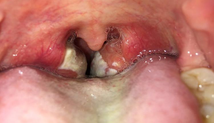
Photos of tonsillitis and pharyngitis
Real images of pharyngeal and tonsillar inflammation caused by viral or bacterial infections.
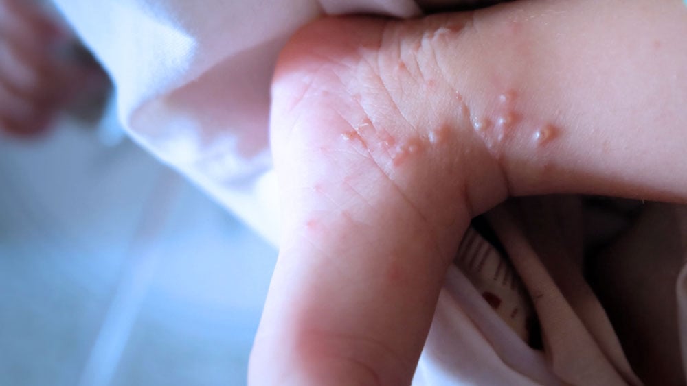
Photos of Dyshidrosis (Dyshidrotic Eczema)
This article presents clinical images of dyshidrosis, illustrating small fluid-filled blisters, usually on the hands and feet, associated with vesicular dermatitis. Photographic analysis aids in the differential diagnosis with mycoses, eczema, and other vesicular dermatoses.
![Pictures: Syphilis – All stages [Warning: Strong Images]](https://www.mdsaude.com/wp-content/uploads/sifilis-imagem.jpg)
Pictures: Syphilis – All stages [Warning: Strong Images]
Images of syphilis manifestations: early (chancre) and late (skin lesions, ulcerations, gummas, and deformities).
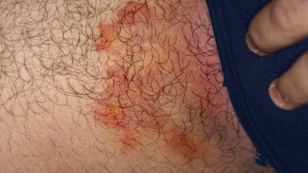
Photos of Jock Itch (Tinea Cruris or Groin Ringworm)
Photos of jock itch (tinea cruris) help identify typical skin lesions, such as reddish patches with rounded edges. The images are useful for educational purposes and to support the clinical diagnosis of fungal infection.
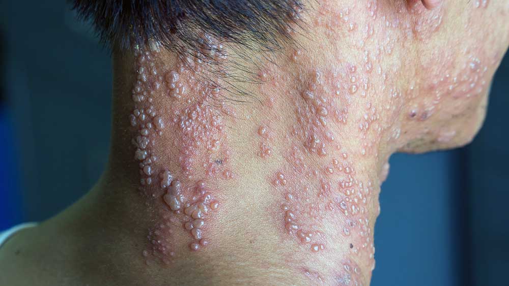
Photos of shingles (herpes zoster)
See real images of shingles (herpes zoster) on different parts of the body. Identify the visual signs of varicella-zoster virus infection and understand how it manifests itself on the skin in a clear and educational way.