

Key facts about erysipelas
Erysipelas is a bacterial skin infection caused mainly by Streptococcus pyogenes. It affects the more superficial layers of the dermis and the lymphatic vessels.
The typical lesion is a red, warm, swollen, and painful area, usually with well-defined borders. Fever, chills, and general malaise may also occur.
The legs are the most commonly affected site. Wounds, athlete’s foot, ulcers, and other skin lesions can provide an entry point for bacteria. Chronic edema, venous insufficiency, and obesity increase the risk.
Erysipelas is not considered a contagious disease and, in most cases, is curable with appropriate antibiotic treatment.
Most patients can be treated with oral antibiotics, but more severe cases may require hospital admission and intravenous antibiotics.
What is erysipelas?
Erysipelas is an acute bacterial skin infection that typically appears as a very red, warm, swollen, and painful area. The lesion usually forms a slightly raised plaque with relatively sharp borders separating it from the surrounding healthy skin. Fever, chills, and general malaise may also occur and, in some patients, develop even before the skin changes become apparent.
The disease is caused mainly by beta-hemolytic streptococci, especially the bacterium Streptococcus pyogenes. The infection primarily affects the superficial portion of the dermis and the superficial cutaneous lymphatic vessels, a feature that helps distinguish it from cellulitis, which tends to involve deeper tissues.
Erysipelas most often affects the lower limbs. The face is another classic location, but any part of the body can be affected. The disease can occur at any age, although it is more likely in people with conditions that facilitate bacterial entry or spread, such as chronic leg edema, venous insufficiency, obesity, diabetes, eczema, ulcers, and other skin lesions.
Although erysipelas is a bacterial infection, it is not considered a contagious disease. In other words, erysipelas itself is not usually transmitted directly from one person with the condition to another. This does not mean that the streptococci responsible for the disease cannot spread between people; rather, erysipelas itself is not usually transmitted from person to person.
How does erysipelas develop?
The skin is the largest organ in the human body and acts as an important barrier between the body and the environment. In addition to preventing excessive water loss and protecting against physical and chemical injury, it makes it more difficult for bacteria, fungi, viruses, and other potentially harmful agents to enter the body. Without an intact skin barrier, deeper tissues would be much more exposed to infection.
At the same time, the surface of the skin is home to a large community of bacteria, fungi, and viruses that make up the skin microbiota. Most of these microorganisms coexist with us without causing disease, and some help maintain the skin barrier and prevent colonization by pathogenic microorganisms.
For erysipelas to develop, there usually needs to be a break in the skin barrier that allows bacteria to enter. This entry point may be obvious, such as a wound, ulcer, or surgical incision, but it is often small and goes unnoticed.
In the lower limbs, common entry points include cracks and maceration between the toes caused by athlete’s foot (tinea pedis). Cuts, abrasions, insect bites, eczema, venous stasis ulcers, and other skin lesions can also allow streptococci to enter. In some patients, no entry point can be identified.
Once the bacteria cross the skin barrier, they multiply in the superficial dermis and reach the local lymphatic vessels, triggering an intense inflammatory response. This produces the characteristic signs of redness, warmth, swelling, and pain.
Which layer of the skin does erysipelas affect?
Several types of infection can affect the skin and adjacent tissues. Examples include fungal skin infections, folliculitis, impetigo, boils, erysipelas, cellulitis, ecthyma, abscesses, and, in even deeper tissue planes, necrotizing fasciitis and osteomyelitis.
These skin infections can be distinguished, among other features, by the anatomical structures they predominantly affect.
Look at the illustration below showing the layers of the skin. Impetigo is a very superficial infection, mainly limited to the epidermis. Erysipelas occurs just below this level, primarily affecting the upper dermis and superficial lymphatic vessels. Cellulitis, on the other hand, involves the deeper dermis and subcutaneous tissue.
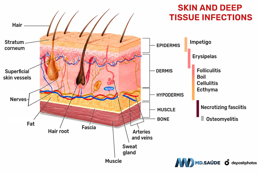
This difference in depth helps explain why erysipelas usually forms a raised plaque with sharper borders, whereas cellulitis tends to produce flatter redness with a less distinct transition to healthy skin. In practice, however, the distinction is not always clear-cut: erysipelas and cellulitis can overlap, and some authors consider erysipelas to be a superficial form of cellulitis.
The depth of the infection helps define which syndrome is present, but it does not determine severity on its own. The extent of the infection, the bacterium involved, the speed of progression, the presence of systemic symptoms, and the patient’s underlying medical conditions are also important.
Differences between erysipelas and cellulitis
Erysipelas and cellulitis are very similar bacterial skin infections. In clinical practice, distinguishing between them is not always easy, and there is some overlap between the definitions used by different authors. In general, however, erysipelas is a more superficial infection with greater involvement of the skin’s lymphatic vessels, whereas cellulitis affects deeper tissues.
The terms cellulitis and cellulite can sometimes cause confusion. Cellulite refers to the cosmetic condition characterized by dimpling and unevenness of the skin surface, particularly in women. This condition, medically known as gynoid lipodystrophy, is not an infection and has no relationship to cellulitis.
Both erysipelas and cellulitis cause redness, warmth, swelling, and pain in the affected area. Fever, chills, and general malaise can also occur with either infection.
The main visible difference is usually found at the edges of the lesion. Because erysipelas is more superficial, the inflamed area tends to be more raised, bright red, and sharply demarcated. In many cases, it is possible to clearly see where the inflamed skin ends and healthy skin begins.
In cellulitis, the inflammation is usually more diffuse. The borders are less distinct, and the transition between the red area and normal skin can be difficult to define. The affected area may also show induration of the subcutaneous tissue.
The image below shows two cases in which these differences are particularly evident.
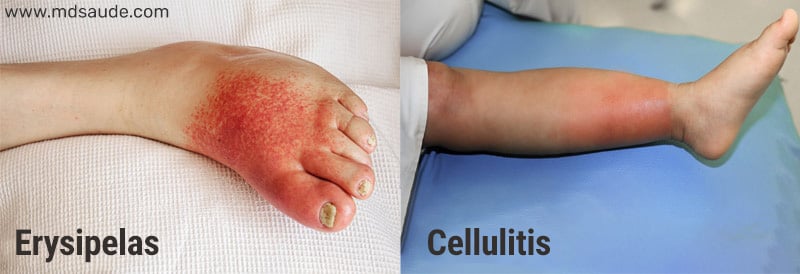
In clinical practice, however, lesions do not always have such typical characteristics. In some cases, erysipelas and cellulitis cannot be reliably distinguished by the appearance of the skin alone. In these situations, treatment is chosen to cover the most likely bacterial pathogens.
The table below summarizes the main differences between erysipelas and cellulitis.
| Erysipelas | Cellulitis | |
|---|---|---|
| Depth of infection | Mainly affects the superficial dermis and superficial cutaneous lymphatic vessels. | Affects the deeper dermis and subcutaneous tissue. |
| Appearance of the lesion | Bright red, warm, painful, and swollen area, often raised and with well-defined borders. Blisters may develop. | Red, warm, painful, and swollen area, generally more diffuse, with less distinct borders and possible tissue induration. |
| General symptoms | Fever, chills, and malaise are common and may develop abruptly, sometimes before the skin lesion becomes apparent. | Fever, chills, and malaise may also occur, especially in more extensive or severe cases. |
| Most common bacteria | Predominantly beta-hemolytic streptococci, especially Streptococcus pyogenes. | Most typical nonpurulent cases are also caused by streptococci. Staphylococcus aureus may be involved, particularly when there is an open wound, penetrating trauma, or an associated purulent infection. |
| Location | Most common on the legs and face but can occur anywhere on the body. | Most common on the legs but can occur anywhere on the body. |
| Complications | May cause abscesses, necrosis, bacteremia, sepsis, and lymphatic damage, particularly when the infection is severe or inadequately treated. | May also lead to abscesses, bacteremia, sepsis, and spread of the infection into deeper tissues. |
As an interesting clinical point, when a facial infection extends onto the pinna of the ear, this finding favors erysipelas over cellulitis. This is known as Milian’s ear sign, because the cartilaginous parts of the external ear contain very little subcutaneous tissue. The sign can help distinguish the two infections, but it does not mean that every red ear is caused by erysipelas, since several other conditions can also cause inflammation in this area.
Risk factors
The presence of an entry point in the skin is one of the main risk factors for erysipelas. In the lower limbs, cracks and maceration between the toes caused by athlete’s foot are particularly important, but any condition that disrupts the skin barrier can facilitate bacterial entry.
Possible entry points include:
- Cuts, abrasions, and wounds.
- Athlete’s foot (tinea pedis), particularly when it causes cracks between the toes.
- Eczema, impetigo, chickenpox, and other conditions that cause skin lesions or intense itching.
- Inflamed or manipulated acne lesions and other small breaks in the skin.
- Mosquito bites and other insect bites.
- Ingrown toenails, onychomycosis, paronychia, and other injuries to the skin around the nails.
- Burns.
- Animal bites.
- Piercings.
- Surgical incisions, injections, and intravenous drug use.
It is important to note that most people who sustain a minor skin injury do not develop erysipelas. This is because disruption of the skin barrier is only part of the problem. Abnormalities in venous or lymphatic circulation and certain medical conditions can also facilitate the development of infection.
The factors most clearly associated with erysipelas, particularly in the lower limbs, include:
- Obesity.
- Chronic ulcers and other skin diseases that impair the skin barrier.
- A previous episode of erysipelas, particularly when edema or lymphatic vessel damage persists.
- Chronic edema of the lower limbs.
- Lymphedema.
- Venous insufficiency of the lower limbs.
Surgical procedures that impair lymphatic drainage, such as lymph node removal, can also increase the risk of erysipelas in the affected limb.
Diabetes mellitus, immunosuppression — including prolonged corticosteroid use — peripheral arterial disease, and excessive alcohol consumption have also been associated with erysipelas in some studies. However, these associations are less consistent than those seen with edema, lymphedema, venous insufficiency, and disruption of the skin barrier.
Bacteria that cause erysipelas
Erysipelas is almost always caused by beta-hemolytic streptococci. The main causative organism is Streptococcus pyogenes, also known as group A beta-hemolytic Streptococcus (GAS).
When there is an entry point in the skin, streptococci can reach the superficial dermis and lymphatic vessels, giving rise to erysipelas.
Although Streptococcus pyogenes is the best-known causative organism, other streptococci can also cause erysipelas, particularly beta-hemolytic streptococci from groups C and G.
Group B Streptococcus (Streptococcus agalactiae) is a less common cause but is important in certain situations. It can cause erysipelas in newborns and, less commonly, may be involved in infections of the abdominal or perineal region in women during the postpartum period.
Other bacteria are much less frequent causes. In particular, there is little evidence that Staphylococcus aureus is an important cause of typical erysipelas, unlike in several other bacterial skin infections.
Symptoms of erysipelas
Erysipelas is almost always unilateral. The lower limbs are involved in approximately 70 to 80% of cases, but the infection can also occur on the face, arms, and other areas of the body.
The typical lesion is a very red, swollen, warm, painful area of skin that is tender to the touch. The borders are usually well defined, making the transition between inflamed and healthy skin relatively easy to identify. Edema and induration can make the skin appear tight, shiny, and, in some cases, give it an “orange-peel” appearance.
Because erysipelas also involves the superficial lymphatic vessels, lymphangitis and enlargement of regional lymph nodes may occur. When the infection affects a leg, for example, the lymph nodes in the groin may become enlarged and painful.
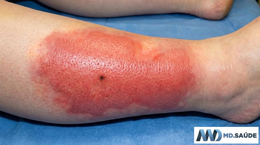
Fever, chills, sweating, and general malaise are common and may begin several hours before the characteristic skin changes appear. Other possible symptoms include headache, loss of appetite, nausea, and occasionally vomiting.
Bullous erysipelas
In some patients, inflammation and skin edema are severe enough to cause blisters, resulting in a form known as bullous erysipelas. The blisters may contain clear fluid or, in more severe cases, blood.
The presence of blisters does not necessarily mean that the infection is deep. However, extensive or hemorrhagic blisters, necrosis, pain that is disproportionate to the appearance of the skin, or rapid progression of the lesion warrant careful medical assessment to rule out more serious forms of infection.
Signs of severe disease and complications
Very severe pain that is disproportionate to the appearance of the skin, very rapid progression of redness and swelling, reduced sensation, crepitus, necrosis, or significant deterioration in the patient’s overall condition are warning signs of deeper infection, particularly necrotizing fasciitis, which is a medical emergency.
Serious complications of erysipelas are uncommon, particularly when treatment is started early. Possible complications include abscess formation, thrombophlebitis, bacteremia, and sepsis. Less commonly, the infection can spread to nearby structures and cause septic arthritis or bone infection. In exceptional cases, bacteria can spread through the bloodstream and cause infections at distant sites, such as endocarditis.
Repeated episodes can also cause progressive damage to the lymphatic vessels and lead to chronic lymphedema. This can create a vicious cycle: erysipelas damages lymphatic drainage, lymphedema increases the risk of further infections, and each new episode can worsen the edema even more.
In extreme and longstanding cases, chronic lymphedema can progress to a severe form of skin thickening and enlargement known as elephantiasis nostras verrucosa, which is unrelated to the elephantiasis caused by filariasis.
How is erysipelas diagnosed?
The diagnosis of erysipelas is primarily clinical. In typical cases, a doctor can usually recognize the condition based on the characteristics of the lesion, the speed at which the symptoms developed, and the presence of predisposing factors or a possible entry point in the skin.
In classic erysipelas without signs of severe disease, laboratory tests are generally not required to confirm the diagnosis or determine treatment.
When blood tests are performed, an increased white blood cell count (leukocytosis) and elevated inflammatory markers, such as C-reactive protein (CRP), are common. However, these findings are nonspecific and do not confirm erysipelas.
Blood cultures or cultures taken from the lesion are also unnecessary in most cases because they frequently fail to identify the causative bacterium. They may be indicated in specific situations, such as severe infection, suspected sepsis, immunosuppression, or an exposure that raises suspicion for less common bacteria.
Imaging tests are not part of the routine evaluation either. Ultrasound may be useful when there is uncertainty about the presence of an abscess, deep vein thrombosis, or a foreign body. CT or MRI may be required when deeper involvement is suspected, such as necrotizing fasciitis, septic arthritis, or osteomyelitis.
Not every red, warm, swollen leg is caused by erysipelas. Stasis dermatitis due to venous insufficiency, deep vein thrombosis, contact dermatitis, and other inflammatory or infectious conditions can produce a similar appearance. Therefore, atypical presentations or cases that do not progress as expected should be reassessed.
Treatment of erysipelas
Most cases of erysipelas can be treated at home with oral antibiotics. In patients with typical erysipelas, no signs of severe disease, and the ability to take medications normally, treatment should include antibiotics with good activity against beta-hemolytic streptococci, which are the main causative organisms.
Commonly used regimens in adults include:
- Penicillin V potassium 500 mg orally every 6 hours.
- Amoxicillin 875 mg orally every 12 hours.
- Cephalexin 500 mg orally every 6 hours.
- Cefadroxil 500 mg orally every 12 hours or 1 g orally once daily.
These regimens are directed primarily against streptococci. In typical erysipelas, there is no need to routinely add antibiotics targeting resistant bacteria such as methicillin-resistant Staphylococcus aureus (MRSA). Broader coverage may be necessary in specific situations, such as the presence of pus, penetrating trauma, injection drug use, a previous MRSA infection, or other circumstances suggesting less common bacteria.
In uncomplicated cases that respond well to antibiotics, 5 to 7 days of treatment is usually sufficient. Treatment may be extended to 10 to 14 days when the response is slower, the infection is severe, or the patient has underlying conditions that make resolution more difficult.
The need for intravenous antibiotics depends mainly on the severity of the illness. An extensive or rapidly progressive infection, significant deterioration in the patient’s overall condition, low blood pressure, inability to take or absorb oral antibiotics, significant immunosuppression, failure of oral treatment, or suspected deeper infection are situations that may justify intravenous treatment and hospital admission.
Facial erysipelas requires particular attention, especially when the infection is close to the eyes or nose. However, facial involvement alone does not mean that antibiotics must be given intravenously.
In patients with a penicillin allergy, the choice of antibiotic depends on the type of allergic reaction and local patterns of bacterial resistance. Macrolides such as erythromycin or clarithromycin may be used in some situations. Roxithromycin and pristinamycin also have evidence of efficacy against erysipelas, but their availability and use vary between countries.
In addition to antibiotics, rest and elevation of the affected limb help reduce edema and relieve pain. Cold compresses and pain relievers can also be used to reduce discomfort.
In patients with chronic edema of the lower limbs, compression is important for controlling swelling and reducing the risk of recurrent episodes. During the acute episode, compression stockings or another form of compression can be continued or resumed as soon as they are comfortable. If compression causes significant pain, it may need to be temporarily stopped and reintroduced as the inflammation improves.
Symptoms usually begin to improve within the first few days after antibiotics are started. Fever, pain, and malaise may improve before the skin begins to look better. Redness and swelling may take 72 hours or longer to begin clearly subsiding and may persist even after the infection is already under control.
If there is no sign of improvement after 48 to 72 hours, or if the redness is spreading rapidly, pain is increasing, or the patient’s general condition is worsening, medical reassessment is necessary.
Prevention of recurrent erysipelas
In patients with repeated episodes of erysipelas, the first step is to identify and control factors that predispose to new infections, particularly athlete’s foot, cracks between the toes, wounds, eczema, venous insufficiency, and chronic edema or lymphedema.
In patients with chronic leg edema, compression therapy can significantly reduce the risk of further episodes.
When recurrences remain frequent despite control of these factors, long-term antibiotic prophylaxis may be indicated. Low-dose oral penicillin V is one of the most extensively studied options. Another possibility is intramuscular benzathine penicillin G, usually administered every 2 to 4 weeks.
There is no single appropriate duration for all patients. The need to continue prophylaxis should be reassessed periodically, taking into account the frequency of recurrent episodes and the possibility of controlling predisposing factors.
- UpToDate — Acute cellulitis and erysipelas in adults: Treatment. UpToDate
- UpToDate — Cellulitis and skin abscess: Epidemiology, microbiology, clinical manifestations, and diagnosis. UpToDate
- National Institute for Health and Care Excellence (NICE) — Cellulitis and erysipelas: antimicrobial prescribing (NG141). NICE guideline NG141
- Infectious Diseases Society of America (IDSA) — Practice Guidelines for the Diagnosis and Management of Skin and Soft Tissue Infections. IDSA guideline
- Centers for Disease Control and Prevention (CDC) — Clinical Guidance for Group A Streptococcal Cellulitis. Updated in 2025. CDC clinical guidance
- Thomas KS, Crook AM, Nunn AJ, et al. Penicillin to Prevent Recurrent Leg Cellulitis. N Engl J Med. 2013;368:1695-1703. doi:10.1056/NEJMoa1206300. New England Journal of Medicine
- Webb E, Neeman T, Bowden FJ, et al. Compression Therapy to Prevent Recurrent Cellulitis of the Leg. N Engl J Med. 2020;383:630-639. doi:10.1056/NEJMoa1917197. New England Journal of Medicine.
- Images: personal archive and Depositphotos.






Reader questions about this topic
Questions selected by the editor for their relevance to this article.